
A few years ago, magnetic bracelets gained popularity all over the world, and their owners sincerely believed that they were now reliably protected from all kinds of human ailments.
Without disputing whether this is true or false, let us recall that as early as the 18th century the well-known physician Mesmer tried to treat his patients with “magnetic water.” Later, the healing properties of magnets were studied by such luminaries of world medicine as Charcot, Durval, Botkin, and Helmholtz. Today it has been firmly established that in disease the magnetic characteristics of blood change noticeably. Therefore, modern physicians successfully use magnetic fields, for example, to reduce a patient’s pain during surgery or in the treatment of sciatica and hypertension.
 |
Meanwhile, the staff of one of the laboratories of the Brooklyn Medical Center, led by Dr. Raymond Damadian, are confident that magnets will help to recognize and cure in time one of the most mysterious diseases of the century—malignant tumors. In short, cancer. The guarantee of this is a successful series of experiments developed and carried out by Brooklyn physicians.
Everything began when the researchers considered how to apply to medicine a physical phenomenon well known to physicists—nuclear magnetic resonance. Let us briefly recall its essence. The atoms of many substances resemble tiny magnets. If they are supplied with additional energy by means of directed magnetic radiation whose frequency coincides with their own, the atoms absorb the energy of the pulse and begin to resonate like a tuning fork.
At this time the atoms move to a higher energy level. But as soon as the radio pulse disappears, the resonance ceases, and the atoms, returning to their previous state, emit excess energy. This radiation can be “captured” by a radio receiver.
Dr. Damadian and his colleagues decided to try to apply the nuclear magnetic resonance effect to the diagnosis of cancer, since most of the atoms of the elements that make up the human body also possess magnetic properties.
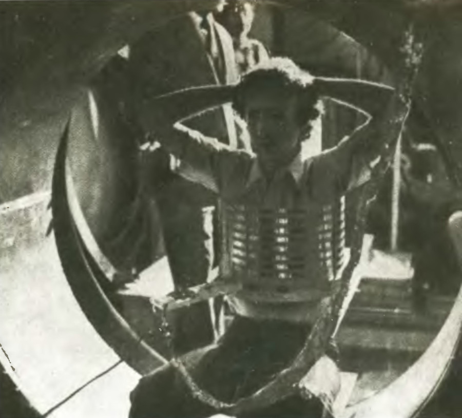
The research was conducted at the intersection of two sciences that at first glance seem far removed from each other—physics and medicine—so the doctor’s laboratory looked unusual. Alongside medical instruments and cages with experimental animals stood complex electronic equipment, a welding machine, a drafting board, tracing paper, and blueprints. Towering above everything was a two-meter ring of complex design. This is the main device with which the scientists are developing a method for the early detection of malignant tumors. Inside the ring—or, more precisely, inside a powerful superconducting magnet capable of generating a uniform field of 1,000 gauss—an adult human can easily fit. The unique structure was designed and built largely by physicians themselves.
They began their experiments with standard electromagnets. To turn them into superconductors, the windings of the electromagnets were cooled with liquid helium (to 4 K). The electromagnets then generated a field with very stable characteristics. Once excited, they required no additional energy.
To induce nuclear magnetic resonance in the first versions of their instruments, the scientists used a special spectrometer that sent radio pulses into a coil containing the object of the experiment. Having received a certain dose of energy, the atoms of the tissues began to resonate. As soon as the supply of radio pulses stopped, they returned to their previous energy level. The energy emitted in this process was captured by the coil and, through amplifiers, fed to the screen of an oscilloscope. The scientists knew that cells of malignant tumors contain about 90% water (healthy tissues about 70%), and that molecules in diseased cells move faster than in normal ones. Consequently, the characteristics of the two types of tissue under nuclear magnetic resonance are completely different.
These differences can be detected by the decay time of the resonance of hydrogen atoms—that is, by the relaxation time. After a series of experiments, the scientists found that in tissues affected by malignant tumors, relaxation occurs almost three times faster than in healthy tissues. These data were obtained in experiments using a short-duration (100 microseconds) radio pulse.
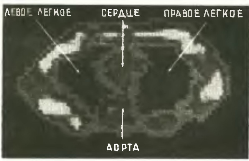 |
| In the photographs: a patient in the magnetic ring; a television image of the chest |
If continuous radio radiation is used within a narrow frequency band (for hydrogen, within 24.1–24.2 MHz), the oscillation frequencies of the atoms of an element will be determined by their position in the molecule. A series of spikes of varying magnitude will appear on the oscilloscope, from which one can judge the chemical structure of the substance under study. This is easy to verify by looking at the oscillograms available in Dr. Damadian’s laboratory. For example, by comparing the resonance of phosphorus atoms in healthy and affected tissues, the scientists found that some of the spikes characteristic of normal tissue are absent in recordings of “diseased” tissue. This, in the opinion of Damadian’s colleagues, is sufficient for the early diagnosis of cancer.
However, as soon as the scientists moved from studying individual tissue samples to a living organism, serious problems arose. In the body, diseased tissues are surrounded by healthy ones, and at first it was almost impossible to separate the signals coming from affected and normal tissues. Another difficulty was that in serial ring magnets with superconducting windings there was room only for small laboratory animals, whereas the goal of Damadian’s experiments was the diagnosis of malignant tumors in humans. Thus, the physicians had to temporarily master the professions of calculator, engineer, and worker.
In order to isolate signals from individual parts of the body, they created a magnetic field focusing system. Now the oscilloscope recorded pulses coming from a strictly limited space whose diameter did not exceed 6 mm. In this way, the researchers learned to excite nuclear magnetic resonance at any point in the human body, thereby determining the chemical composition of the tissue.
Much more effort went into constructing a giant electromagnet. Dr. Damadian and his staff spent a great deal of time on calculations and drawings. But once the project was completed, it turned out that no American company was capable of building such a “ring.” Manufacturers were deterred by the very design of the unique installation: how, for example, could one ensure high precision of the winding elements, whose connections also had to have extremely low resistance? And how could one ensure especially reliable welding of the vessels intended to store liquid nitrogen and helium cooled to ultralow temperatures? Once again, the scientists themselves took up the task. Working 16 hours a day, over five weeks Doctors Goldsmith and Stanford wound 48 km of superconducting magnet coil from titanium–niobium wire. Fortunately, this alloy does not require welding, and all connections could be made using cold methods.
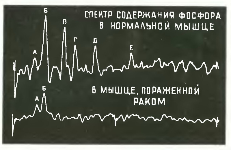 |
| On the graph: comparative phosphorus content in healthy and affected muscles. |
Then the researchers turned to the vessels for liquefied gases. Having mastered the skills of professional welders, after four months they created original electrodes: the standard ones were unsuitable because they did not guarantee the required tightness of the cooling system, which consisted of three concentric vessels. In the first vessel, filled with liquid helium, the magnet itself is located. It, in turn, is enclosed in a second vessel (with liquid nitrogen). All this is placed, as it were, in a casing from which the air has been pumped out. Does this not closely resemble the well-known Dewar flask, only of an unusual shape?
And finally, the installation began to operate, providing a constant, uniform magnetic field of 1,000 gauss. The object or subject under study moves inside the ring in such a way that the focal point fixed at the center of the ring makes, as it were, a transverse slice through the entire depth of the body. At this time, a special coil attached to the subject “captures” the signals of nuclear magnetic resonance. The signals are fed into a computer, which converts them into a conditional color image (the color code is constructed in accordance with the frequency of the incoming signal) that can be seen on a television screen (see figure).
Thus, without a scalpel, physicians penetrate the human body, learning about the chemical composition of tissues. By changes in the intensity of signals from hydrogen atoms, they judge the water content of tissues. If there is unusually much of it, it is time to sound the alarm—this is an unmistakable sign of malignant degeneration of tissues.
In a decisive experiment, the role of the experimental subject fell to laboratory staff member Dr. Minkov. Having secured the pulse generator coil to his chest, he took a seat inside the magnetic ring on a mechanically driven chair. The magnetic field now, as it were, scanned his body in a single plane. On the television screen, colored squares appeared, carrying information about the chemical composition of tissues.
The results of this experiment exceeded the boldest expectations of the participants. The aorta zone, through which blood flowed, appeared on the screen as a distinct orange spot, whereas the lungs filled with air produced a weak signal. On the screen, their area was colored dark blue. If Minkov’s lungs had been affected by cancer, the tumor would have shown an abnormally bright spot.
Despite the success, Dr. Damadian and his colleagues realize that much work still lies ahead.
Nevertheless, it can already be said that Dr. Damadian’s discovery is another step on the path to victory over one of the most terrible diseases. And who knows—perhaps scientists will create magnetic installations generating radiation of such frequency and power that diseased cells will begin to break down, while healthy ones will simply not perceive it. But from experiments with a giant magnet to clinical application, many years of work still remain...